Types Of Hemangioma Ppt
They can grow on the surface or deeper into the center canal of a bone. It is called vascular nevus.
 Vascular Malformations
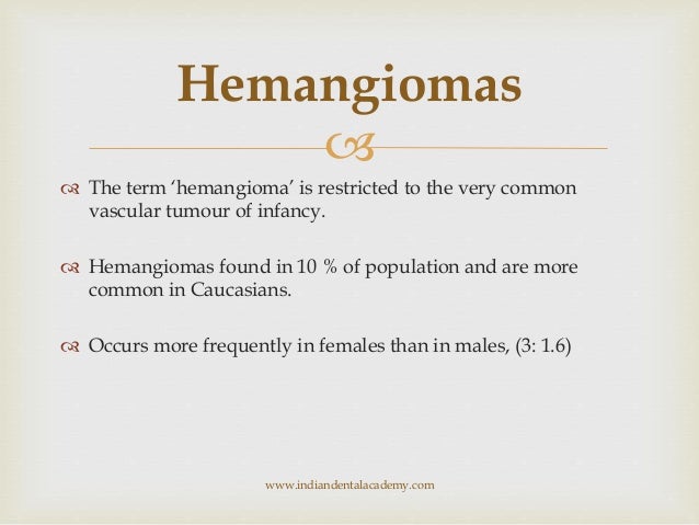
Vertebral hemangiomas are a common etiology estimated to be found in 10-12 of humans at autopsy.
Types of hemangioma ppt. There are three principle types. Doctors may diagnose a congenital hemangioma on a prenatal ultrasound. You can go for beta-blockers laser treatment or surgery to have them removed from your body.
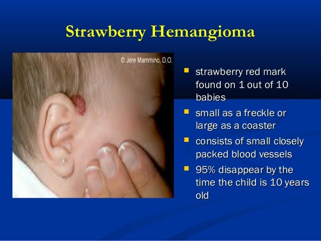
An infantile hemangioma hee-man-jee-OH-muh is a type of birthmark that happens when a tangled group of blood vessels grows in or under a babys skin. All brain tumors are uncommon and brain hemangiomas make up a small proportion of these. Two types of hemangioma occur in the brain.
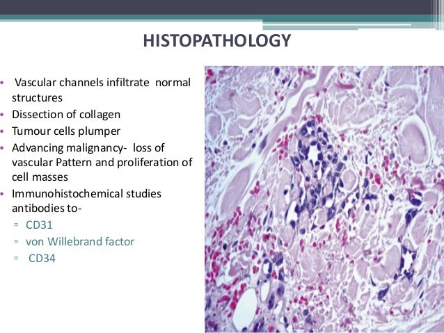
Progenitorstem cells HemSC endothelial cells HemEC and pericytes HemPericytes. Because they typically do not cause symptoms these tumors are often found by chance when an x-ray or MRI image is taken for another purpose. How to Cure Different Types of Hemangiomas - Hemangiomas can be treated in several ways.
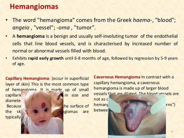
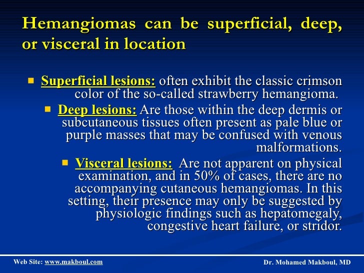
Superficial on the surface of the skin deep under the skin and mixed. Often exhibit the classic crimson color of the so-called strawberry hemangioma. Symptoms if they do occur are usually related to large hemangiomas trauma the hormonal and hemodynamic changes of pregnancy or osseous expansion and extra-osseous.
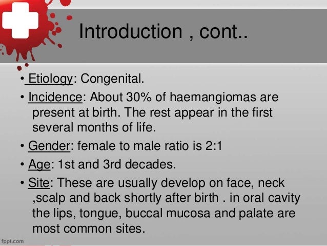
It may be congenital or traumatic in origin. Hemangioma is a skin disorder which occurs due to an unusual accumulation of blood vessels resulting in the formation of red scar on or under the skin surface. Bishop in Gnepps Diagnostic Surgical Pathology of the Head and Neck Third Edition 2021 Clinical Features.
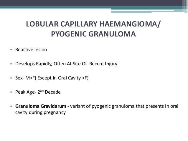
Non-involuting NICH Lobulated capillary hemangiomas LCH pyogenic granuloma Tufted angioma TA Others Locally aggressive Kaposiform hemangioendothelioma KHE Kaposi sarcoma Others Malignant Angiosarcoma Others Adapted from the International Society for the Study of. Commonly these are benign lesions that are found incidentally during radiology studies for other indications. They are benign in nature and frequently asymptomatic.
Shallow on the outside of the skin These take a gander from the outset and afterward become brilliant red with a raised lopsided surface. A hemangioma is a common vascular birthmark made of extra blood vessels in the skin. Hemangiomas are benign tumors of the endothelial cells which normally line.
Infantile hemangioma IH Congenital hemangioma rapidly involuting RICH. Half of the cases of synovial hemangioma represent cavernous lesions rest are examples of capillary hemangioma arteriovenous hemangioma pure venous hemangioma. Blended These hemangiomas have both shallow and.
Uncommon related to the nerve involved and include pain paresthesiae numbness. 8 H HemSC represent a small percentage of proliferating hemangioma cells and have the ability for self-renewal and multilineage differentiation. A congenital hemangioma hem-an-gee-o-ma is a vascular lesion that is present and fully grown at birth.
Infantile hemangiomas become visible in the first few days to weeks after a baby is born. A vertebral hemangioma is a vascular lesion within a vertebral body. Introduction A hemangioma is an abnormal buildup of blood vessels in the skin or internal organs It may be congenital or traumatic in origin It is called vascular nevus composed of seemingly disorganized vessels that are filled with blood and is connected to the main vein.
291 Many capillary hemangiomas present either at birth or immediately thereafter and show an initial period of. A hemangioma is an abnormal build up of blood vessels in the skin or internal organs. Capillary hemangiomas are the most common subtype of hemangiomas and are the most common subtype of soft-tissue tumor in infants and children.
Hemangiomas may happen anyplace on the body. Capillary and cavernous types are the most common hemangiomas found in bone. How to Cure Different Types of Hemangiomas - Hemangiomas can be treated in several ways.
Several cell types have been isolated from hemangiomas. PowerPoint PPT presentation free to view. Before the year 2000 these lesions were grouped in with infantile hemangiomas.
Profound under the skin These show up as a somewhat blue purple growing with a smooth surface. Calcifications or prominent scarring with hyalinization sclerosed hemangioma. A few kids may have mutiple.
Hemangiomas can be superficial deep or visceral in location Superficial lesions. Every treatment has its own pros and cons so discuss each and every option with your doctor. You can go for beta-blockers laser treatment or surgery to have them removed from your body.
Although uncommon hemangiomas can develop in internal organs most often the liver and intestines. There are three main types of hemangioma. Intraneural hemangioma Clinical features.
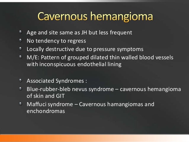
Every treatment has its own pros and cons so discuss each and every option with your doctor. CONGENITAL HEMANGIOMA RICH Rapid Involuting Congenital Hemangioma Red-violaceous color Central telengectasia Peripheral pale halo occasional Superficial ulceration Central nodularity Transient Thrombocytopenia Platelet level increase with regression Mainly seen in trunk extrimities Rapidly involute during first week month NICH Non Involuting Congenital Hemangioma Ovoid macular slightly raised Light gray in colour Prominent coarse telengectasia Warm on palpation Predisposition in. Composed of seemingly disorganized vessels that are filled with blood and is connected to the main vein.
 Vascular Tumors Of Soft Tissue
 Infantile Haemangioma The Lancet
 Hemangioma Online Presentation
 Hemangioma
 Haemangioma
 Haemangioma And Vascular Anomelies
 Haemangiomas And Vascular Malformations
 Hemangioma
 Benign Tumors Tumor Like Lesions Of The Oral Cavity Ppt Download
 Hemangiomas And Vascular Malformations Certified Fixed Orthodontic C
 Infantile Hemangioma
Infantile Hemangioma
 Final Hemangioma
 Infantile Hemangioma
 Final Hemangioma
 Hemangiomas James Hansen Ppt Video Online Download
 Haemangioma And Vascular Anomelies
 Hemangioma
Hemangioma
 Haemangiomas And Vascular Malformations